|
Schmidt MA1, Herman EM2.
BMC Plant Biol. 2018 Dec 13;18(1):354. doi: 10.1186/s12870-018-1579-8.
Abstract
BACKGROUND:
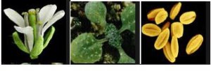
Soybean is a globally important oil seed crop. Both the high protein and oil content of soybean seeds make this crop a lucrative commodity. As in higher eukaryotic species with available genomes, the functional annotation of most of soybean's genes still remains to be investigated. A major hurdle in the functional genomics of soybean is a rapid method to test gene constructs before embarking on stable transformation experiments.
RESULTS:
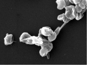
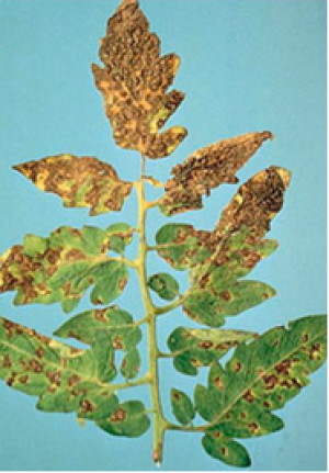
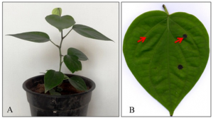
In this paper we describe the morphology and composition of the persistent single-cell aleurone layer that derives from the endosperm of developing soybean seeds. Its composition compared to cotyledonary tissue indicates the aleurone layer plays a role in both abiotic and biotic stress. The potential utility as the aleurone layer as a transient expression system in soybean was shown. As a near transparent single-cell layer it can be used as a transient expression system to study transgene expression and inter- and intra-cellular targeting as it is amenable to microscopic techniques.
CONCLUSION:
The transparent single cell aleurone layer was shown to be compositionally comparable to cotyledonary tissue in soybeanwith an enrichment in oxidative response proteins and shown to be a potential transient expression platform.
See https://www.ncbi.nlm.nih.gov/pubmed/30545296
(19).png "Characterization and functional biology of the soybean aleurone layer.")
Figure 1: Morphological structure of soybean aleurone layer. a) Mature cotyledon showing the isolation of the single-cell aleurone layer from the seed coat. b) Light micrograph of a cross-section of a mature soybean cotyledon showing the single cell layer of the aleurone, crushed endosperm and storage parenchyma cells. Bar denotes 10 μm. c) Transmission electron micrograph of an aleurone layer isolated from a mature soybean cotyledon. Among the intracellular constituents marked are: PSV protein storage vacuole, P plastid, G golgi, M mitochondria, ER endoplasmic reticulum. Circled areas show the connections between adjacent cells where the plasmadesmata are visible. Bar denotes 1 μm
|
[ Other News ]___________________________________________________
|


 Curently online :
12
Curently online :
12 Total visitors :
7515132
Total visitors :
7515132