|

 Curently online :
8 Curently online :
8 |
|
 Total visitors :
7514794 Total visitors :
7514794 |
|
|
Glycosyltransferase MDR1 assembles a dividing ring for mitochondrial proliferation comprising polyglucan nanofilaments
Saturday, 2017/12/23 | 06:36:13
|
|
Yamato Yoshida, Haruko Kuroiwa, Takashi Shimada, Masaki Yoshida, Mio Ohnuma, Takayuki Fujiwara, Yuuta Imoto, Fumi Yagisawa, Keiji Nishida, Shunsuke Hirooka, Osami Misumi, Yuko Mogi, Yoshihiko Akakabe, Kazunobu Matsushita, and Tsuneyoshi Kuroiwa PNAS December 12 2017; vol.114; no.50: 13284–13289 SignificanceThe mitochondrion-dividing (MD) ring mediates binary division of mitochondria. However, the molecular identity of the MD ring is currently unknown. We show that the glycosyltransferase MITOCHONDRION-DIVIDING RING1 (MDR1) regulates the synthesis of the polyglucan nanofilament bundle that assembles the MD ring. MDR1 is essential for mitochondrial division and forms a single ring at the mitochondrial division site in the unicellular red alga Cyanidioschyzon merolae. Nanoscale imaging and componential analysis demonstrated that MDR1 is involved in MD ring formation and that the MD ring filaments are composed of polymeric-glucose nanofilaments. An MDR1 homologue performs a similar function in chloroplast division, suggesting that the establishment of the MDR1 family was crucial for the emergence of endosymbiotic organelles. AbstractMitochondria, which evolved from a free-living bacterial ancestor, contain their own genomes and genetic systems and are produced from preexisting mitochondria by binary division. The mitochondrion-dividing (MD) ring is the main skeletal structure of the mitochondrial division machinery. However, the assembly mechanism and molecular identity of the MD ring are unknown. Multi-omics analysis of isolated mitochondrial division machinery from the unicellular alga Cyanidioschyzon merolae revealed an uncharacterized glycosyltransferase, MITOCHONDRION-DIVIDING RING1 (MDR1), which is specifically expressed during mitochondrial division and forms a single ring at the mitochondrial division site. Nanoscale imaging using immunoelectron microscopy and componential analysis demonstrated that MDR1 is involved in MD ring formation and that the MD ring filaments are composed of glycosylated MDR1 and polymeric glucose nanofilaments. Down-regulation of MDR1 strongly interrupted mitochondrial division and obstructed MD ring assembly. Taken together, our results suggest that MDR1 mediates the synthesis of polyglucan nanofilaments that assemble to form the MD ring. Given that a homolog of MDR1 performs similar functions in chloroplast division, the establishment of MDR1 family proteins appears to have been a singular, crucial event for the emergence of endosymbiotic organelles.
See: http://www.pnas.org/content/114/50/13284.full
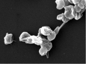
Fig. 1. Identification and expression profiles of the mitochondrial division gene MDR1 via multi-omics analysis. (A) Phase contrast and immunofluorescence images of interphase (Left) and dividing (Right) cells of the unicellular red alga C. merolae. The mitochondrial division machinery and the mitochondria were immunostained with anti-Mda1 antibodies (Mda1, yellow) and antimitochondrial porin antibodies (mito, red). (Scale bar: 1 μm.) (B) Schematic representation of organelle- and cell-division processes of C. merolae. MD, mitochondrial division machinery; mt, mitochondrion; n, nucleus; PD, plastid division machinery; pt, plastid. (C and D) EM images of dividing C. merolae cells. The Inset in C shows the mitochondrial division site (boxed area in C). The MD ring (white arrowheads) was observed as electron-dense deposits on the top and bottom of a dividing mitochondrion (C) and was visualized by saccharide staining in early (D, Upper) and late mitochondrial division phases (D, Lower). (Scale bars: 200 nm in C and D; 100 nm in Inset.) (E) Immunofluorescence and EM images of isolated division machinery complexes containing mitochondrial division machinery (green) and plastid division machinery (red). The Inset shows a schematic representation of the mitochondrial (green) and plastid (red) division machinery in the EM image. (Scale bars: 500 nm, Left; 200 nm, Right.) (F and G) Mascot score histograms (F) and pie chart (G) of the 185 identified genes in the fraction of isolated division machinery complexes. (See Table S1.) (H, Left) Hierarchical clustering analysis of gene expression patterns of the identified genes using the time-course transcriptome dataset. (H, Right) The two groups that contain known endosymbiotic organelle division genes and exhibit specific expression patterns. (See Fig. S3.) (I) Domain architecture of CMJ262C/MDR1. A specific motif in the glycosyltransferase domain is visualized. (See Fig. S4.) (J and K) Immunoblot analysis of the protein expression profiles of MDR1 using anti-MDR1 antibody (J) in synchronized cell cultures (K). IB, immunoblot. (L and M) Comparison of the amounts of MDR1 protein at each isolation step. Twenty micrograms of protein per sample were loaded onto each lane. P, pellet; IB, immunoblot; S, supernatant. Lane 1, whole cell; lane 2, isolated mitochondria and plastid; lane 3, isolated mitochondrial and plastid membranes; lane 4, isolated division machinery complexes. |
|
|
|
[ Other News ]___________________________________________________
|
(47).png "Glycosyltransferase MDR1 assembles a dividing ring for mitochondrial proliferation comprising polyglucan nanofilaments")