|

 Curently online :
6 Curently online :
6 |
|
 Total visitors :
7517271 Total visitors :
7517271 |
|
|
Identification of MEDIATOR16 as the Arabidopsis COBRA suppressor MONGOOSE1
Saturday, 2016/01/02 | 16:37:36
|
|
Nadav Sorek, Heidi Szemenyei, Hagit Sorek, Abigail Landers, Heather Knight, Stefan Bauer, David E. Wemmer, and Chris R. Somerville
Significance
The cobra mutants of Arabidopsis, such as cob-6, have impaired growth associated with a defect in cellulose synthesis. Mutations in MEDIATOR16 (MED16) reduce the number of misregulated genes in cob-6 mutants and suppress the phenotypes. This observation implicates MED16 in transcriptional responses to cell wall defects. Ectopic expression of two pectin methylesterase inhibitors (PMEIs) identified in a suppressor screen partially suppressed the growth defect in the cob-6 mutant. The results confirm that the PMEIs have significant in vivo activity, and provide evidence that pectin esterification can modulate cell wall properties.
Abstract
We performed a screen for genetic suppressors of cobra, an Arabidopsis mutant with defects in cellulose formation and an increased ratio of unesterified/esterified pectin. We identified a suppressor named mongoose1 (mon1) that suppressed the growth defects of cobra, partially restored cellulose levels, and restored the esterification ratio of pectin to wild-type levels. mon1 was mapped to the MEDIATOR16 (MED16) locus, a tail mediator subunit, also known as SENSITIVE TO FREEZING6 (SFR6). When separated from the cobra mutation, mutations in MED16 caused resistance to cellulose biosynthesis inhibitors, consistent with their ability to suppress the cobra cellulose deficiency. Transcriptome analysis revealed that a number of cell wall genes are misregulated in med16 mutants. Two of these genes encode pectin methylesterase inhibitors, which, when ectopically expressed, partially suppressed the cobra phenotype. This suggests that cellulose biosynthesis can be affected by the esterification levels of pectin, possibly through modifying cell wall integrity or the interaction of pectin and cellulose.
See: http://www.pnas.org/content/112/52/16048.abstract.html?etoc
PNAS December 29, 2015 vol. 112 no. 52: 16048–16053
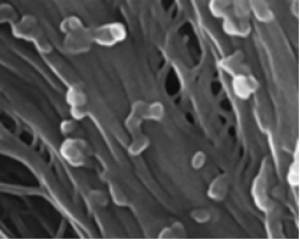
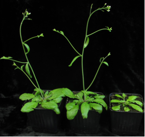
Fig. 2. Cellulose macrostructure and amount in mon1 cob-6 mutant. (A) Cellulose in root cells stained with S4B. In cob-6, staining is reduced and less homogeneous, with some cells exhibiting almost a complete lack of fluorescence. Fibrils that can be detected are not as defined as in wild type, and are not as regularly oriented (Upper, cob-6). In mon1 cob-6, fibrils were similar to wild type, more so in elongated cells. (Scale bars, 15 μm.) (B) One-dimensional ssNMR analysis. Quantitative 13C direct polarization (DP)-MAS ssNMR spectra of wild-type, cob-6, and mon1 cob-6 cell walls. Additional annotation is shown in Fig. S8. (C) Relative intensities of interior and surface cellulose C4 signals from 13C DP-MAS spectra. |
|
|
|
[ Other News ]___________________________________________________
|
.gif "Identification of MEDIATOR16 as the Arabidopsis COBRA suppressor MONGOOSE1")