|

 Curently online :
6 Curently online :
6 |
|
 Total visitors :
7479680 Total visitors :
7479680 |
|
|
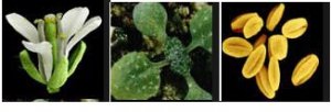
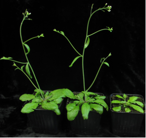
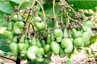
Insect haptoelectrical stimulation of Venus flytrap triggers exocytosis in gland cells
Sunday, 2017/05/07 | 06:16:56
|
|
Sönke Scherzer, Lana Shabala, Benjamin Hedrich, Jörg Fromm, Hubert Bauer, Eberhard Munz, Peter Jakob, Khaled A. S. Al-Rascheid, Ines Kreuzer, Dirk Becker, Monika Eiblmeier, Heinz Rennenberg, Sergey Shabala, Malcolm Bennett, Erwin Neher, and Rainer Hedrich SignificanceThe Venus flytrap has been in the focus of scientists since Darwin’s time. Carnivorous plants, with their specialized lifestyle, including insect capture, as well as digestion and absorption of prey, developed unique tools to gain scarce nutrients. In this study, we describe mechanistic insights into the cascade of events following the capture of insect prey. Action potentials evoked by the struggling prey are translated into touch-inducible hormone signals that promote the formation of secretory vesicles. Different varieties of digestive compounds are released sequentially into the flytrap’s “green stomach” and break down the captured animal. Amperometry provides insight into the kinetics and chemistry of the stimulus-coupled glandular secretion process. AbstractThe Venus flytrap Dionaea muscipula captures insects and consumes their flesh. Prey contacting touch-sensitive hairs trigger traveling electrical waves. These action potentials (APs) cause rapid closure of the trap and activate secretory functions of glands, which cover its inner surface. Such prey-induced haptoelectric stimulation activates the touch hormone jasmonate (JA) signaling pathway, which initiates secretion of an acidic hydrolase mixture to decompose the victim and acquire the animal nutrients. Although postulated since Darwin’s pioneering studies, these secretory events have not been recorded so far. Using advanced analytical and imaging techniques, such as vibrating ion-selective electrodes, carbon fiber amperometry, and magnetic resonance imaging, we monitored stimulus-coupled glandular secretion into the flytrap. Trigger-hair bending or direct application of JA caused a quantal release of oxidizable material from gland cells monitored as distinct amperometric spikes. Spikes reminiscent of exocytotic events in secretory animal cells progressively increased in frequency, reaching steady state 1 d after stimulation. Our data indicate that trigger-hair mechanical stimulation evokes APs. Gland cells translate APs into touch-inducible JA signaling that promotes the formation of secretory vesicles. Early vesicles loaded with H+ and Cl− fuse with the plasma membrane, hyperacidifying the “green stomach”-like digestive organ, whereas subsequent ones carry hydrolases and nutrient transporters, together with a glutathione redox moiety, which is likely to act as the major detected compound in amperometry. Hence, when glands perceive the haptoelectrical stimulation, secretory vesicles are tailored to be released in a sequence that optimizes digestion of the captured animal.
See: http://www.pnas.org/content/114/18/4822.abstract.html?etoc PNAS May 2 2017; vol.114; no.18: 4822–4827
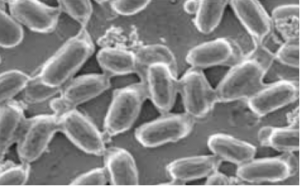
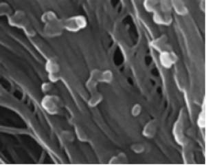
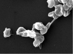
Fig. 1. Exocytotic vesicle fusion is stimulated in activated gland complexes. EMs of the outer layer of resting (A and C) and COR-stimulated (B, D, and E) Dionaea gland complexes are shown. A detailed view (A, B, and E) and overview (C and D) are shown. Whereas resting glands only exhibit a few exocytotic events, a massive rise in exocytotic vesicle fusion with the plasma membrane (black arrows) could be detected 48 h after COR stimulation. B, dark-stained body; C, cuticle; CW, cell wall; ER, endoplasmic reticulum; M, mitochondria; S, secreted fluid; V, vacuole. Slight shadow lines are due to carrier film handling during TEM sample preparation; all images are noncomposite originals. |
|
|
|
[ Other News ]___________________________________________________
|
(22).png "Insect haptoelectrical stimulation of Venus flytrap triggers exocytosis in gland cells")