|

 Curently online :
9 Curently online :
9 |
|
 Total visitors :
7517636 Total visitors :
7517636 |
|
|
Multiple functional self-association interfaces in plant TIR domains
Saturday, 2017/03/11 | 06:05:23
|
|
Xiaoxiao Zhang, Maud Bernoux, Adam R. Bentham, Toby E. Newman, Thomas Ve, Lachlan W. Casey, Tom M. Raaymakers, Jian Hu, Tristan I. Croll, Karl J. Schreiber, Brian J. Staskawicz, Peter A. Anderson, Kee Hoon Sohn, Simon J. Williams, Peter N. Dodds, and Bostjan Kobe SignificanceAutophagy contributes to innate immune responses in metazoans by targeted elimination of intracellular pathogens, including viruses, in a process termed “xenophagy.” Whether autophagy has a similar role in plant immunity is unknown. Here we demonstrate that the selective autophagy receptor NEIGHBOR OF BRCA1 (NBR1) binds the viral capsid protein and particles of cauliflower mosaic virus (CaMV) and mediates their autophagic degradation. We further demonstrate that this antiviral xenophagy is counteracted by protective functions of autophagy-resistant CaMV inclusion bodies. Finally, we show that a second, nonselective NBR1-independent autophagy pathway promotes plant viability during infection and serves as a proviral mechanism to extend the timespan for virus production and potential CaMV transmission. Thus autophagy exhibits important pro- and antiviral roles in compatible plant–virus interactions. AbstractAutophagy plays a paramount role in mammalian antiviral immunity including direct targeting of viruses and their individual components, and many viruses have evolved measures to antagonize or even exploit autophagy mechanisms for the benefit of infection. In plants, however, the functions of autophagy in host immunity and viral pathogenesis are poorly understood. In this study, we have identified both anti- and proviral roles of autophagy in the compatible interaction of cauliflower mosaic virus (CaMV), a double-stranded DNA pararetrovirus, with the model plant Arabidopsis thaliana. We show that the autophagy cargo receptor NEIGHBOR OF BRCA1 (NBR1) targets nonassembled and virus particle-forming capsid proteins to mediate their autophagy-dependent degradation, thereby restricting the establishment of CaMV infection. Intriguingly, the CaMV-induced virus factory inclusions seem to protect against autophagic destruction by sequestering capsid proteins and coordinating particle assembly and storage. In addition, we found that virus-triggered autophagy prevents extensive senescence and tissue death of infected plants in a largely NBR1-independent manner. This survival function significantly extends the timespan of virus production, thereby increasing the chances for virus particle acquisition by aphid vectors and CaMV transmission. Together, our results provide evidence for the integration of selective autophagy into plant immunity against viruses and reveal potential viral strategies to evade and adapt autophagic processes for successful pathogenesis.
See http://www.pnas.org/content/114/10/E2026.abstract.html?etoc PNAS Narch 7 2017; vol.114; no.10: E2026–E2035
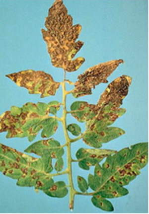
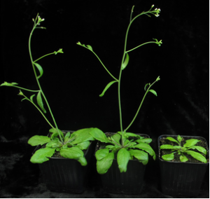
Fig. 1. Autophagy promotes plant fitness and suppresses CaMV accumulation. (A) Virus-induced symptoms in WT, atg5, atg7, atg5 npr1, npr1, and nbr1 plants at 28 dai with CaMV strain CM1841 (Lower Row) compared with noninfected controls (Upper Row). (Scale bar, 20 mm.) (B) Ratio of total chlorophyll content in infected and noninfected plants. Error bars represent SD (n = 6). (C) CaMV capsid protein (CP; P4) levels determined by ELISA in systemic leaves of the indicated genotypes at 14 dai. Values are shown as means ± SD (n = 4) and are presented as arbitrary units relative to WT plants. (D) CaMV DNA levels determined by qPCR in systemic leaves of WT, atg5, and nbr1 plants at 14 dai. Values represent means ± SD (n = 4) relative to WT plants and were normalized with 18S ribosomal DNA as the internal reference. (E) CaMV RNA levels determined by RT-qPCR as an individual 35S transcript or via the leader sequence as the sum of 35S, 19S, and 8S transcripts in systemic leaves of WT, atg5, and nbr1 plants at 14 dai. Values are shown as means ± SD (n = 3 biological replicates) relative to the WT 35S transcript level. (F) Immunoblot analysis of CaMV P2, P3, P4, and P6 accumulation in WT, atg5, and nbr1 plants. Total proteins were extracted from whole plants at 14 dai and probed with specific antibodies. Noninfected WT plants served as control (C) for signal background, and Ponceau S (PS) staining verified comparable protein loading. Statistical significance (*P < 0.05; **P < 0.01) was revealed by Student´s t test (compared with WT). |
|
|
|
[ Other News ]___________________________________________________
|
(45).png "Multiple functional self-association interfaces in plant TIR domains")