|

 Curently online :
9 Curently online :
9 |
|
 Total visitors :
7514586 Total visitors :
7514586 |
|
|
Phytophthora palmivora establishes tissue-specific intracellular infection structures in the earliest divergent land plant lineage
Saturday, 2018/04/28 | 07:42:01
|
|
Philip Carella, Anna Gogleva, Marta Tomaselli, Carolin Alfs and Sebastian Schornack PNAS April 17, 2018. Vol. 115 (16): E3846-E3855 SignificanceDespite the importance of liverworts as the earliest diverging land plant lineage to support fungal symbiosis, it is unknown whether filamentous pathogens can establish intracellular interactions within living cells of these nonvascular plants. Here, we demonstrate that an oomycete pathogen invades Marchantia polymorpha and related liverworts to form intracellular infection structures inside cells of the photosynthetic layer. Plants lacking this tissue layer display enhanced resistance to infection, revealing an architectural susceptibility factor in complex thalloid liverworts. Moreover, we show that dedicated host cellular trafficking proteins are recruited to pathogen interfaces within liverwort cells, supporting the idea that intracellular responses to microbial invasion originated in nonvascular plants. AbstractThe expansion of plants onto land was a formative event that brought forth profound changes to the earth’s geochemistry and biota. Filamentous eukaryotic microbes developed the ability to colonize plant tissues early during the evolution of land plants, as demonstrated by intimate, symbiosis-like associations in >400 million-year-old fossils. However, the degree to which filamentous microbes establish pathogenic interactions with early divergent land plants is unclear. Here, we demonstrate that the broad host-range oomycete pathogen Phytophthora palmivora colonizes liverworts, the earliest divergent land plant lineage. We show that P. palmivora establishes a complex tissue-specific interaction with Marchantia polymorpha, where it completes a full infection cycle within air chambers of the dorsal photosynthetic layer. Remarkably, P. palmivora invaginates M. polymorpha cells with haustoria-like structures that accumulate host cellular trafficking machinery and the membrane syntaxin MpSYP13B, but not the related MpSYP13A. Our results indicate that the intracellular accommodation of filamentous microbes is an ancient plant trait that is successfully exploited by pathogens like P. palmivora.
See: http://www.pnas.org/content/115/16/E3846
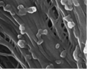
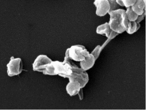
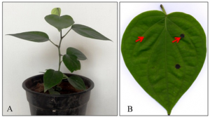
Figure 1: P. palmivora colonizes the photosynthetic layer of M. polymorpha. (A) Disease symptoms of 3-wk-old M. polymorpha TAK1 (male) thalli inoculated with P. palmivora ARI-td zoospores or water (Mock) over a 7-d time course. (B) Epifluorescence microscopy demonstrating the spread of P. palmivora growth across TAK1 thalli from 1 to 4 dpi. Epifluorescence (Epifluor.) from the pathogen is displayed alongside bright-field (BF) images. (Scale bars, 500 μm.) (C) Confocal fluorescence microscopy of sectioned TAK1 thalli infected with P. palmivora at 7 dpi. Z-stack projections of red fluorescence from the pathogen are displayed alone (Left, tdTomato) or merged with all channels (Right, bright-field and plastid autofluorescence in turquoise). Arrowheads indicate air pores. (Scale bars, 100 μm.) (D) Cryo-SEM image of TAK1 thalli colonized by P. palmivora at 7 dpi. Mechanically fractured air chamber demonstrating hyphal growth within the chamber (yellow arrows) and sporangia (Sp) at the air pore. (E) Cryo-SEM image showing intercellular (yellow arrows) and intracellular (red arrow) associations between P. palmivora hyphae and photosynthetic filaments within M. polymorpha air chambers at 7 dpi. (Scale bars, 20 μm.) All experiments were performed at least three times, with similar results. |
|
|
|
[ Other News ]___________________________________________________
|
(49).png "Phytophthora palmivora establishes tissue-specific intracellular infection structures in the earliest divergent land plant lineage")