|

 Curently online :
10 Curently online :
10 |
|
 Total visitors :
7471219 Total visitors :
7471219 |
|
|
Two independent S-phase checkpoints regulate appressorium-mediated plant infection by the rice blast fungus Magnaporthe oryzae
Thursday, 2017/01/12 | 07:53:31
|
|
Míriam Osés-Ruiz, Wasin Sakulkoo, George R. Littlejohn, Magdalena Martin-Urdiroz, and Nicholas J. Talbot SignificanceRice blast is a devastating fungal disease of cultivated rice, and its control is vital to ensure global food security. In an effort to understand how the rice blast fungus causes disease, we have investigated how the cell cycle controls the early stages of plant infection. The rice blast fungus develops a special cell, called an appressorium, to infect rice leaves. This structure generates enormous pressure, which the fungus applies as physical force to puncture the leaf surface. We have shown that a buildup of pressure in the appressorium is necessary to trigger an unusual cell-cycle checkpoint that is necessary for the appressorium to function properly. If this process is blocked, rice blast disease cannot occur. AbstractTo cause rice blast disease, the fungal pathogen Magnaporthe oryzae develops a specialized infection structure called an appressorium. This dome-shaped, melanin-pigmented cell generates enormous turgor and applies physical force to rupture the rice leaf cuticle using a rigid penetration peg. Appressorium-mediated infection requires septin-dependent reorientation of the F-actin cytoskeleton at the base of the infection cell, which organizes polarity determinants necessary for plant cell invasion. Here, we show that plant infection by M. oryzae requires two independent S-phase cell-cycle checkpoints. Initial formation of appressoria on the rice leaf surface requires an S-phase checkpoint that acts through the DNA damage response (DDR) pathway, involving the Cds1 kinase. By contrast, appressorium repolarization involves a novel, DDR-independent S-phase checkpoint, triggered by appressorium turgor generation and melanization. This second checkpoint specifically regulates septin-dependent, NADPH oxidase-regulated F-actin dynamics to organize the appressorium pore and facilitate entry of the fungus into host tissue.
See: http://www.pnas.org/content/114/2/E237.abstract.html?etoc PNAS January 10, 2016; vol.114; no.2: E237–E244
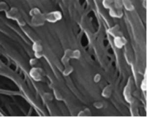
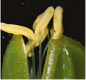
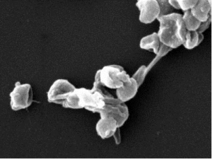
Fig. 1. Appressorium morphogenesis is an S-phase-regulated developmental process in M. oryzae. (A) Micrographs to show effect of inhibition of DNA replication by 200 mM HU on appressorium formation in M. oryzae, added at 2 h postinoculation (hpi) and observed at 24 h. (Scale bar, 20 µm.) (B) Bar chart to show frequency of appressorium formation after exposure to HU. ****P < 0.0001 (unpaired Student’s t test; n = 3 experiments; spores = 300). (C) Diagram of LacO/LacI operator system. A construct containing 256 LacO repeats was integrated at a random locus in the genome. Fluorescence was visualized by expressing GFP–LacI–NLS, which binds to LacO repeats. In a prereplicative cell (G1) in which a single locus is present, a single punctum is observed, whereas in a postreplicative (S/G2) nucleus, two puncta appear. (Scale bar, 10 µm.) (D) Micrographs to show appressorium formation of Guy11, nim1I327E, cyc1nimE10, cyc1nimE6, and bim1F1673* mutants at 24 °C and 30 °C. (Scale bar, 20 µm.) (E) Bar chart to show frequency of appressorium formation by Guy11, nim1I327E, cyc1nimE10, cyc1nimE6, and bim1F1673* mutants at 24 and 30 °C. **P < 0.01; ***P < 0.001; ****P < 0.0001 (unpaired Student’s t test; n = 3 experiments; spores observed = 300). |
|
|
|
[ Other News ]___________________________________________________
|
.png "Two independent S-phase checkpoints regulate appressorium-mediated plant infection by the rice blast fungus Magnaporthe oryzae")