|

 Curently online :
35 Curently online :
35 |
|
 Total visitors :
7659364 Total visitors :
7659364 |
|
|
A peptide-mediated, multilateral molecular dialogue for the coordination of pollen wall formation
Friday, 2022/06/10 | 08:24:48
|
|
Jekaterina Truskina, Stefanie Brück, Annick Stintzi, Sophy Boeuf, Satoshi Fujita, Niko Geldner, Andreas Schaller, Nicolas M. Doll, and Gwyneth C. Ingram PNAS May 24, 2022; 119 (22) e2201446119 SignificancePollen viability depends on a tough external barrier called the pollen wall. Pollen wall components are produced by tapetum cells, which surround developing pollen grains within the anther. Precise coordination of tapetum activity with pollen grain development is required to ensure effective pollen wall formation. Here, we reveal that this is achieved through a multidirectional dialogue involving three distinct cell types. We show that peptide precursors from the tapetum are activated by proteases produced stage specifically in developing pollen grains. Unexpectedly, we found that activated peptides are perceived not in the tapetum, but in the middle layer, which encloses the developing tapetum and pollen grains, revealing an unsuspected role for this enigmatic cell layer in the control of tapetum development. AbstractThe surface of pollen grains is reinforced by pollen wall components produced noncell autonomously by tapetum cells that surround developing pollen within the male floral organ, the anther. Here, we show that tapetum activity is regulated by the GASSHO (GSO) receptor-like kinase pathway, controlled by two sulfated peptides, CASPARIAN STRIP INTEGRITY FACTOR 3 (CIF3) and CIF4, the precursors of which are expressed in the tapetum itself. Coordination of tapetum activity with pollen grain development depends on the action of subtilases, including AtSBT5.4, which are produced stage specifically by developing pollen grains. Tapetum-derived CIF precursors are processed by subtilases, triggering GSO-dependent tapetum activation. We show that the GSO receptors act from the middle layer, a tissue surrounding the tapetum and developing pollen. Three concentrically organized cell types, therefore, cooperate to coordinate pollen wall deposition through a multilateral molecular dialogue.
See https://www.pnas.org/doi/10.1073/pnas.2201446119
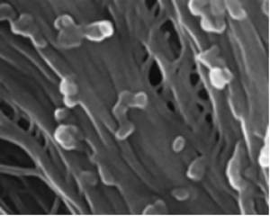
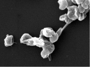
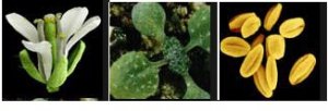
Figure 1: GSO1 and GSO2 RLKs are required for pollen wall formation and tapetum development. (A) Inside the anther, the haploid (n) pollen are surrounded by four diploid (2n) sporophytic cell layers: tapetum, middle layer, endothecium, and epidermis. (B) At the end of the tetrad stage (stage 7), the tapetum cells start to synthesize and release sporopollenin precursors into the locular matrix, where they ultimately attach to the surface of the pollen grains. When pollen wall construction is finished (stage 11), the tapetum cells degrade, and the resulting cellular debris also adheres to the pollen surface. Pollen wall structural components are indicated. The exine is composed of nexine, bacula, and tectum. (C–F) SEM images of the wild type (C and D) and the gso1-1 gso2-1 (E and F) mature pollen. (G and H) Pollen and anther development in wild-type anthers (G) and gso1-1 gso2-1 anthers (H) from the tetrad stage (stage 7) to pollen release (stage 13). The anther cross-sections are stained with acriflavine. (I–L) Pollen wall formation in wild-type (I and J) and the gso1-1 gso2-1 mutant (K and L) anthers shown by toluidine blue staining at stages 8 (I and K) and 10 (J and L). Black arrow indicates reddish-purple staining (toluidine blue) on the surface of the pollen grains in the gso1-1 gso2-1 mutant; red arrow indicates ectopic yellow color detected between tapetum cells in the gso1-1 gso2-1 mutant. (M–P) TEM of the pollen wall in wild-type (M and N) and gso1-1 gso2-1 (O and P) pollen. Arrow indicates fused pollen wall in the mutant. (Q and R) Cryo-SEM of the pollen wall in wild-type (Q) and gso1-1 gso2-1 (R) pollen. Arrows indicate the pollen walls. (S and T) Pollen wall material stained with Auramine-O in wild-type (S) and gso1-1 gso2-1 (T) anthers. Arrows indicate ectopic staining in the mutant. (U and V) TEM showing ectopic deposition of sporopollenin-like material around the tapetum cells in the gso1-1 gso2-1 mutant (V) (arrow) compared to the wild type (U). Scale bars: C and E, 50 µm; G and H, 20 µm; D, F, I–L, S, and T, 10 µm; M–R, U, and V, 1 µm. P, pollen; T, tapetum; ML, middle layer.
|
|
|
|
[ Other News ]___________________________________________________
|
(204).png "A peptide-mediated, multilateral molecular dialogue for the coordination of pollen wall formation")