|
Yu Zhou, Xiao-Ming Xia, and Christopher J. Lingle
Subject: PHYSIOLOGY
Significance
The voltage- and Ca2+-activated BK-type (BK) K+ channels serve as a key regulator of electrical and Ca2+ signaling as well as a model system for studying allosteric regulation of protein. By examining Cd2+ coordination with cysteines placed in BK S6 inner helix, our work reveals a previously undefined structural feature in the BK S6 at a position critical for the coupling between voltage sensor activation and channel opening. In addition, we show that BK S6 does not form an ion permeation gate in its cytosolic end. These results define specific structural constraints in the BK inner pore region that differ from voltage-dependent K+ (Kv) channels.
Abstract
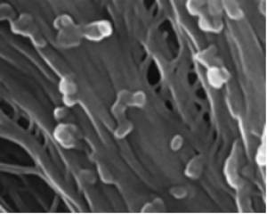
To probe structure and gating-associated conformational changes in BK-type potassium (BK) channels, we examined consequences of Cd2+ coordination with cysteines introduced at two positions in the BK inner pore. At V319C, the equivalent of valine in the conserved Kv proline-valine-proline (PVP) motif, Cd2+ forms intrasubunit coordination with a native glutamate E321, which would place the side chains of V319C and E321 much closer together than observed in voltage-dependent K+ (Kv) channel structures, requiring that the proline between V319C and E321 introduces a kink in the BK S6 inner helix sharper than that observed in Kv channel structures. At inner pore position A316C, Cd2+ binds with modest state dependence, suggesting the absence of an ion permeation gate at the cytosolic side of BK channel. These results highlight fundamental structural differences between BK and Kv channels in their inner pore region, which likely underlie differences in voltage-dependent gating between these channels.
See: http://www.pnas.org/content/112/16/5237.abstract.html?etoc
PNAS April 21, 2015 vol. 112 no. 16 5237-5242
(15).png "Cadmium–cysteine coordination in the BK inner pore region and its structural and functional implications")
Fig. 5. A hypothetic structure of the BK PGD based on the Kv1.2 crystal structure and functional results from our previous and current studies. (A) The hypothetic structure of BK PGD (orange) is superimposed with the Kv1.2 structure (blue). Three pore-lining residues of BK channel (A313, A316, and S317) are colored in green. The S5 of the BK structure is omitted for clarity. K+ ions are rendered as purple balls. Lipid membrane is delimited by red dotted lines. The boxed region is magnified in B to show the structure around the YVP/PVP motif. Side chains of BK319, BK321, Kv1.2406, and Kv1.2408 (equivalent to Shaker474 and Shaker476) are rendered as ball and chain. Notice that Kv1.2406 and Kv1.2408 are not on the same surface of the Kv S6 helix.
|
[ Other News ]___________________________________________________
|

 Curently online :
36
Curently online :
36 Total visitors :
7732231
Total visitors :
7732231