|

 Curently online :
62 Curently online :
62 |
|
 Total visitors :
7654924 Total visitors :
7654924 |
|
|
Quantitative proteomics reveals common and unique molecular mechanisms underlying beneficial effects of caffeine and trigonelline on human hepatocytes
Sunday, 2023/03/12 | 07:08:30
|
|
Paleerath Peerapen, Chanettee Chanthick, Visith Thongboonkerd Biomed Pharmacother.; 2023 Feb;158:114124. doi: 10.1016/j.biopha.2022.114124. AbstractCaffeine and trigonelline are the major bioactive compounds in coffee. Caffeine alone or combined with other coffee compounds shows hepatoprotective effects. However, molecular mechanisms underlying such hepatoprotective effects remain unclear. We therefore addressed molecular effects of caffeine and trigonelline on human hepatocytes using quantitative proteomics followed by bioinformatic analyses to obtain topological and functional significance. HepG2 cells were treated with 100 μM caffeine or trigonelline for 24-h and evaluated by quantitative proteomics using nanoLC-ESI-LTQ-Orbitrap MS/MS. A total of 26 and 25 significantly altered proteins were identified in caffeine-treated and trigonelline-treated cells, respectively, compared with control cells. Topological analyses revealed that ribosomal and translation regulatory proteins predominantly served as the hub proteins associated with protein clusters. Functional analyses also revealed that these two bioactive compounds shared some molecular mechanisms via induction of translational processes. There were also other unique molecular functions and biological processes triggered or suppressed by either caffeine or trigonelline. These data highlight common and unique molecular mechanisms underlying the hepatoprotective effects of caffeine and trigonelline that may be useful for future clinical applications.
See https://pubmed.ncbi.nlm.nih.gov/36521247/
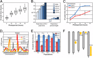
Fig. 1. : Screening for the optimal concentration, quantitative proteomics data and validation. (A): HepG2 cells were treated with 0.1, 1, 10, 100, or 1000 μM caffeine or trigonelline for 24-h, whereas the untreated cells served as the control. Percentage of cell death was then analyzed. No significant difference was detected. (B): Venn diagram summarizing numbers of significantly altered proteins in caffeine-treated and trigonelline-treated HepG2 cells. (C) and (D): Western blot analysis to confirm upregulations of HSP70 induced by caffeine and HSP90 induced by trigonelline. GAPDH (glyceraldehyde-3-phosphate dehydrogenase) served as the loading control. The full-length images of all biological replicates of these blots are provided in Supplementary Fig. S3. All of these quantitative data were obtained from three independent experiments using independent biological samples. The data shown in (A) and (D) are reported as mean ± SEM. |
|
|
|
[ Other News ]___________________________________________________
|
(123).png "Quantitative proteomics reveals common and unique molecular mechanisms underlying beneficial effects of caffeine and trigonelline on human hepatocytes")